New Pulmonary Hypertension Mapping Technology Finds Dysfunction in Left Ventricle


Right ventricular dysfunction is intimately linked to pulmonary hypertension (PH). Since the dynamics of the two ventricles of the heart are connected through the shared tissue of the septum – the wall between the two ventricles – the team behind a new British study hypothesized that the left ventricle would also be affected in PH, and aimed to try a new mapping technology to study their theory.
The study, “Left ventricular diastolic dysfunction in pulmonary hypertension predicts functional capacity and clinical worsening: a tissue phase mapping study,“ was recently published in the Journal of Cardiovascular Magnetic Resonance.
Daniel S. Knight and colleagues from the UCL Institute of Cardiovascular Science in London used a novel cardiovascular magnetic resonance (CMR) tissue phase mapping (TPM) technique to assess left ventricle myocardial function in patients with PH.
The method differs from other CMR techniques by analyzing velocity in multiple planes, rather than the strain of the myocardium.
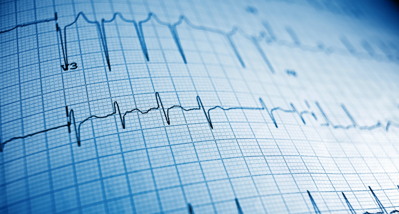
The team enrolled 40 PH patients and 20 healthy controls and investigated their diastolic function, especially focusing on the velocity and shape of the E-wave on the electrocardiogram. The E-wave, or early diastolic wave, is a measure of the rush of blood flowing into the left ventricle as soon as the cardiac valve opens.
Researchers had known that the left ventricle diastolic function was abnormal in PH patients. Hypothesizing that the TPM results could be linked to functional capacity, the participants underwent a six-minute walk distance (6-MWD) test. The team found that one particular parameter, peak radial E-wave velocity, was predictive of functional capacity.
In addition, an association between another parameter – peak global longitudinal E-wave velocity – and clinical worsening was also observed.
The results indicated that the mechanics of left ventricle function are indeed affected by the high pressure in the right ventricle. The TPM technique can, therefore, be used for detecting illness and predicting clinical outcome. In fact, the technique might be superior to other methods in identifying clinical worsening in PH patients.
The authors argue that left-ventricle pathology might actively contribute to symptoms in PH. Since the method is new, the team believes the study needs to be expanded with more patients who are followed over a longer period of time to verify the usefulness of the technique. If the method becomes established in clinical practice, it could also be used to assess the effects of vasodilator drugs in PH.