
Philips Improves Diagnostic Capabilities of Its Echocardiogram System
Written by |
Philips recently announced two new capabilities of its echocardiogram system, the EPIQ CVx cardiology ultrasound platform, that will assist in heart imaging and functional studies, improving the accuracy and duplication of diagnostic tests for pulmonary hypertension (PH), among other conditions.
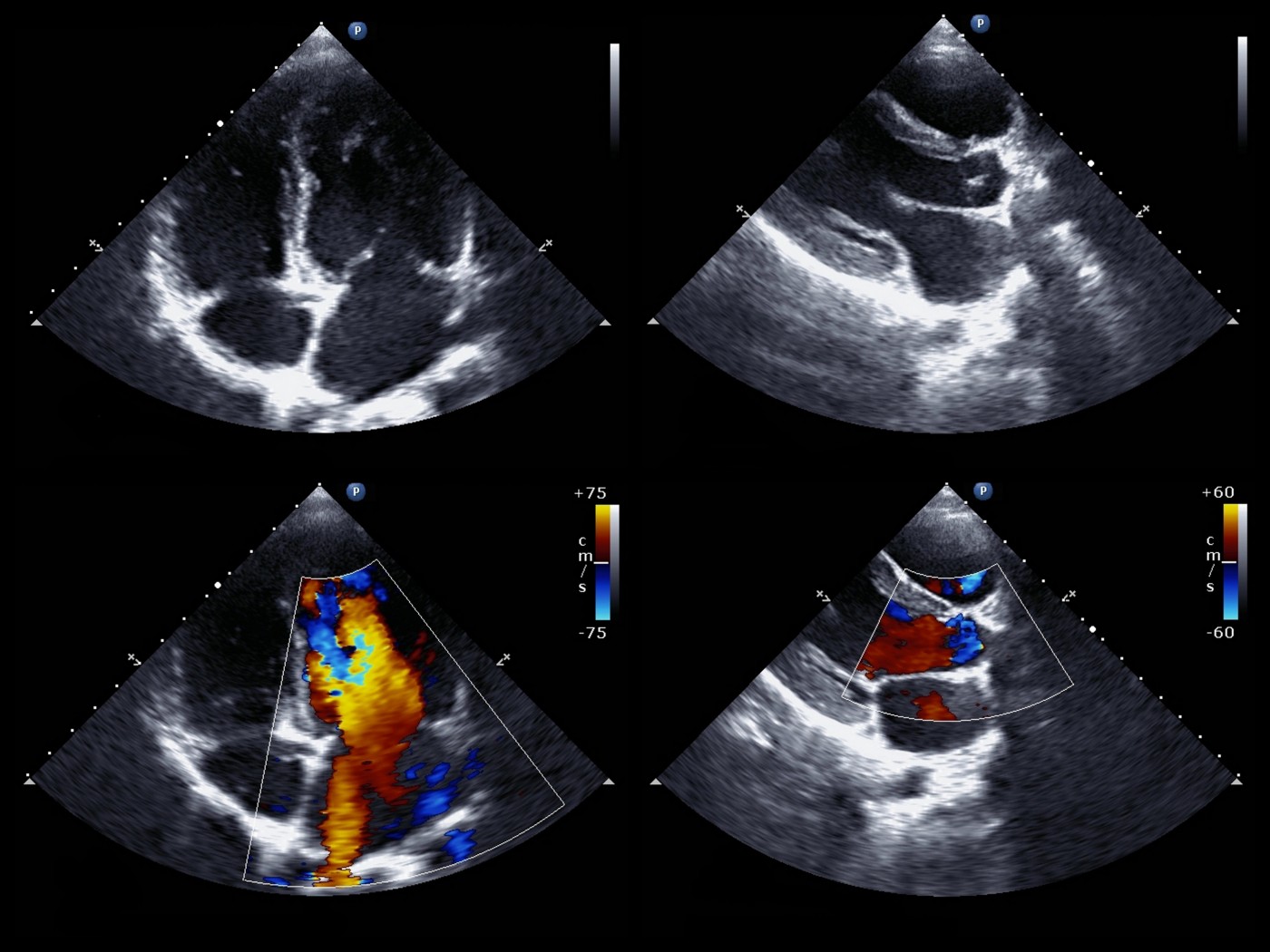
The system improvements are the result of new automation software, called the EPIQ CVx Release 5.0. It provides 2D visualization of the heart and a 3D assessment of the volume of blood in two lower chambers of the heart (ventricles) and of the amount of blood leaving the heart with every contraction (ejection fraction).
“The complexity of cardiac exams can be a barrier to accessing high-quality care. Philips is addressing this by leveraging Artificial Intelligence to make echo exams easier, faster and more reproducible,” David Handler, general manager of cardiac ultrasound at Philips, said in a press release.
Both ventricle volume and ejection fraction are vital features for PH diagnosis and for understanding PH progression.
“By incorporating advanced automation, there is less variability between scans, leading to accurate treatment decisions which benefits patients,” Handler said.
The automation application also results in more accurate assessment of heart function with fewer scans than previously required, thereby limiting the need for frequent use of the equipment. According to Handler, “The new release of EPIQ CVx is a major step forward, reducing the number of touches of the system by 21% in each exam, which is equivalent to more than 400 exams each year.”
The EPIQ CVx Release 5.0 includes applications that measure the strain exerted on the heart tissues in three chambers of the heart, including the left ventricle (AutoStrain LV), right ventricle (AutoStrain RV), and the left atrium (AutoStrain LA). The Autostrain applications use advanced automatic view recognition technology that provides the exceptional visualization of heart muscles and help in the analysis of their function.
The Release 5.0 also has the 3D Auto RV application, which was developed to provide clear images of the heart’s right ventricle, enabling accurate quantification of cardiac function in as little as 15 seconds, the press release stated.
In addition to these capabilities, the update also extends the use of this echocardiogram platform to the field of cardio-oncology. With the new updates, echocardiography can now be used to accurately monitor heart health during chemotherapy.
These new functional capabilities of EPIQ CVx cardiology ultrasound platform add to its existing feature called the Dynamic HeartModel, which provides clear images of the left side of the heart and its function — the region where heart failure usually begins.
The complete feature of EPIQ CVx Release 5.0, along with its diagnostic and interventional guide, were presented at the 2019 annual meeting of the American Society of Echocardiography (ASE) in Portland, Oregon.


Leave a comment
Fill in the required fields to post. Your email address will not be published.