New Accurate, Time-Saving Heart Visualization Method Aids Physicians in Treating PAH, Heart Disease Patients


Ventripoint Diagnostics‘ VMS, a new heart visualization system for cardiac MRI and 3D ultrasounds, was recently deemed an accurate method by a German group of researchers, the company announced on Monday.
According to information available on Ventripoint’s website, VMS was originally designed for congenital heart disease, with subsequent clearance for Pulmonary Arterial Hypertension and non-specific heart disease in both Canada and Europe, although FDA clearance is still pending for PAH.
[adrotate group=”4″]
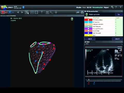
The system is used to evaluate right heart function, using a knowledge-based reconstruction (KBR), which is a library of specific hearts. Ultrasound images are acquired using standard clinical views, allowing the physician or sonographer to identify anatomical landmarks with dots on a number of the 2D ultrasound views throughout the heart. The system uses a sensor on the ultrasound transducer to track where the 2D slice plane is in 3D space, which provides precise anatomical landmark location.
The study, led by Dr. Kai Laser and published in the Journal of the American Society of Ecocardiography, was motivated by the fact that right ventricular volume quantification using real-time three-dimensional echocardiographic (RT3DE) imaging is limited by technical shortcomings of acquisition and quantification. Thus, the VMS offers potentially promising technology for better imaging, diagnosing, and treatment of those with PAH and heart disease.
Researchers used a two-step approach to overcome these limitations, first applying a modified acquisition technique for RT3DE imaging, and secondly using a software tool that used knowledge-based reconstruction (the method used by Ventripoint’s VMS system).
[adrotate group=”3″]
For the study, sixty individuals, of which 20 were healthy and 40 had congenital heart defects, consecutively underwent investigation by cardiac magnetic resonance and RT3DE imaging. CMR data sets were first quantified by the method of disks as the standard, and then CMR and RT3DE data sets were quantified using the knowledge-based reconstruction software and compared with the method of disks.
Results showed that intraobserver variability was excellent for KBR of CMR and RT3DE data, with he KBR analysis requiring a mean time of 5 minutes. The team thus classified the KBR as an accurate, versatile, and time-saving method for right ventricular three-dimensional volumetry, suggesting that the VMS system is clinically valuable.
Dr. George Adams, CEO of Ventripoint, stated in a press release that this study “hows how the VMS technology can be applied across multiple imaging modalities and provides accurate and equivalent results for Right Ventricular function,” adding that the system’s ability “greatly increases” the company’s market potential, as it allows them to seek partners who would integrate the VMS approach into MRI and 3D-echocardiography analysis packages, while continuing to market VMS for routine 2D-echocardiography.”