Estrogen May Help Protect Against Heart Changes in PAH, Animal Study Suggests


Estrogen, one of the major female sex hormones, has a protective effect in pulmonary arterial hypertension (PAH) by halting oxidative stress and collagen deposition, a rat study suggests.
The study, “Effect of estrogen on right ventricular remodeling of monocrotaline-induced pulmonary arterial hypertension in rats and its mechanism,” was published in the journal European Review for Medical and Pharmacological Sciences.
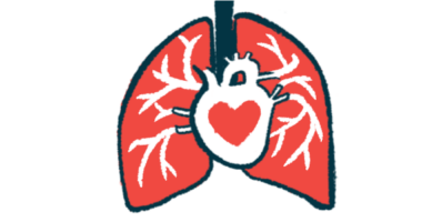
In pulmonary hypertension, lung arteries are contracted or blocked, resulting in an increased blood pressure that makes the heart’s right ventricle work harder to maintain a proper blood flow into the lungs. This vascular resistance is inevitably accompanied by a remodeling of the right ventricle, which may lead to respiratory failure, pulmonary heart disease, heart failure, and eventually death.
Previous studies suggested that oxidative stress induces right ventricle remodeling, mediated by an enzyme called NOX4. Oxidative stress is caused by an imbalance between the body’s production of potentially harmful reactive oxygen species and its ability to contain them.
PAH has been reported to occur more in females than males, particularly in young women. This led researchers to ask whether estrogen — one of the two major sex hormones in women — could play a role in this discrepancy between males and females.
Interested in PH research? Check out our forums and join the conversation!
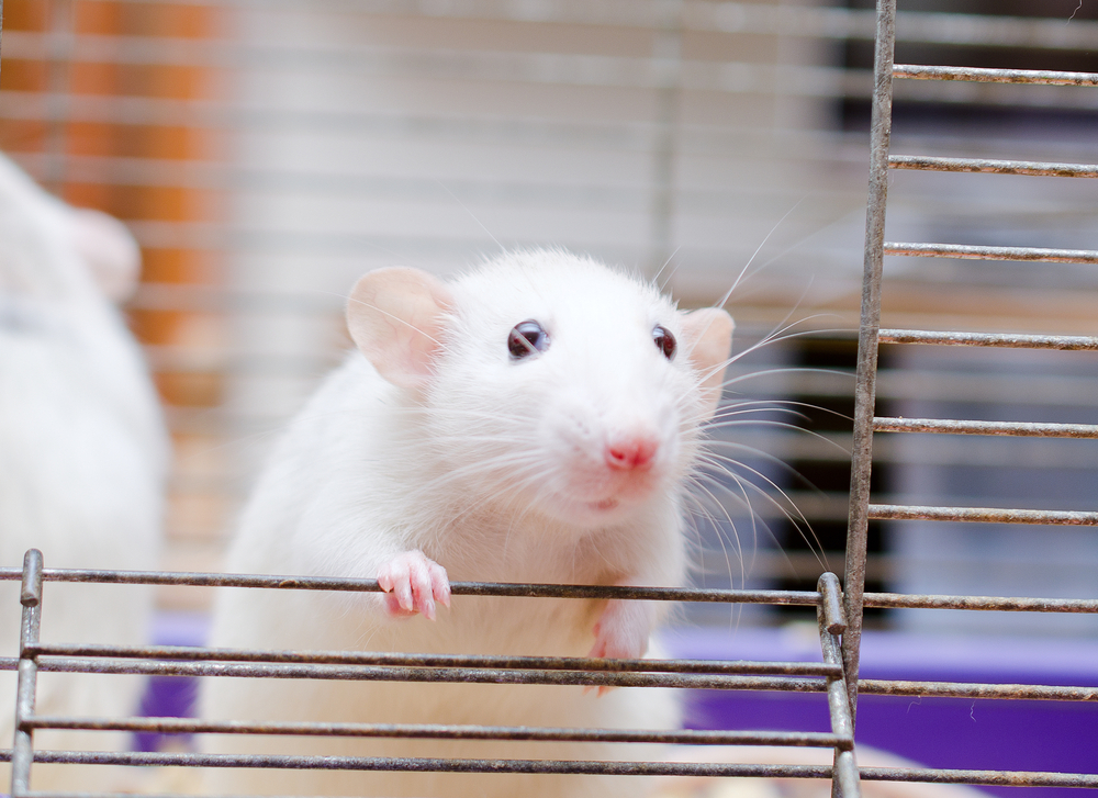
To address this question, they used rats with chemically induced PAH via a compound called monocrotaline. The animals were given estrogen as a compound called 17-β estradiol. They tested two different estradiol doses: 50 and 100 milligrams per kilogram a day, injected into the skin, over four weeks.
Results showed that rats with chemically induced PAH developed the disease, shown by a significant increase in pulmonary arterial pressure. Moreover, the animals presented hypertrophy of the right ventricle, meaning that the ventricle was enlarged due to the effort necessary to compensate for the higher pulmonary arterial blood pressure, similar to what happens in PAH patients.
Treatment with either dose of estrogen led to a significant decrease in both right ventricular systolic pressure (RVSP) and mean pulmonary arterial pressure (mPAP), supporting the inhibitory role of estrogen on ventricle remodeling and suggesting that it could improve the hemodynamic (blood flow) parameters in PAH rats.
Histology analysis of heart tissue showed that cardiomyocytes, the heart muscle cells, were enlarged and disorganized in PAH rats. However, these tissue lesions were significantly reduced with 17-β estradiol treatment.
Further histological tests showed that estrogen treatments also reduced the deposition of extracellular matrix, namely the collagen protein that is normally increased in PAH.
Next, the team performed a molecular study to understand the mechanism behind estrogen’s protective effect. They observed that the levels of NOX4 (an enzyme involved in oxidative stress) decreased in PAH rats after treatment with 17-β estradiol. Additionally, the activity of the NF-κB transcription factor — activated in PAH and promoting collagen production — was also reduced in the right ventricle of estrogen-treated rats.
These results suggest that estrogen plays a role in PAH. The team suggested that by acting on oxidative stress regulation and collagen deposition, estrogen decreases the right ventricle remodeling characteristic of PAH.
“We found that the 17-β estradiol could remarkably alleviate MCT [monocrotaline]-induced right ventricular remodeling of PAH rats. It is suggested that 17-β estradiol exerts its protective role in PAH by inhibiting NOX4-mediated oxidative stress and NF-κB-mediated collagen deposition,” the team concluded.