Pulmonary hypertension diagnosis
Last updated Oct. 24, 2025, by Lindsey Shapiro, PhD

Reaching a pulmonary hypertension (PH) diagnosis can be challenging because its symptoms develop slowly and overlap with those of other heart and lung conditions.
PH is characterized by elevated pressure in the pulmonary arteries, which carry blood from the heart to the lungs. Doctors will carefully evaluate a person’s medical history and run several diagnostic tests to confirm a diagnosis of PH and determine its underlying cause.
The earlier an accurate PH diagnosis is established, the sooner appropriate PH treatment can be initiated, which may help slow disease progression and ease symptoms.
Initial steps
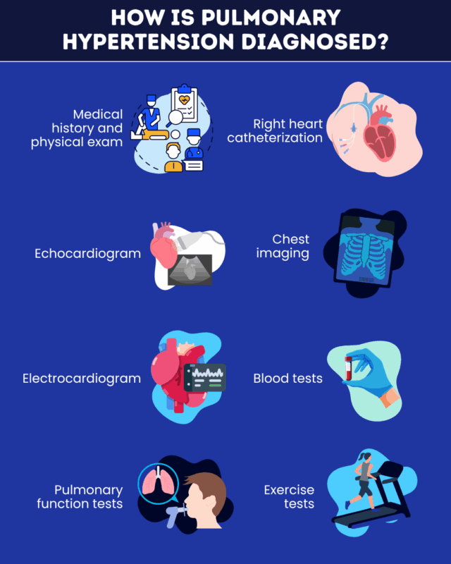
Physicians usually start the diagnostic process by collecting a thorough medical history and performing a physical exam. They’ll ask about common PH symptoms such as:
- shortness of breath (dyspnea)
- excessive fatigue
- dizziness or fainting
- chest pressure or pain
- irregular heartbeat
- swelling in the legs, ankles, or abdomen
During the physical exam, they’ll listen to the heart and lungs, check blood oxygen levels, and look for other signs of PH.
They’ll also ask about risk factors for PH, such as:
- medication use
- family history of PH
- any other existing medical issues
PH diagnostic tests
If a doctor suspects PH based on the initial exam, they’ll still need to run other PH screening tests to confirm it.
Per PH diagnosis criteria, the disease can be diagnosed when tests indicate elevated pulmonary artery pressure above a certain threshold.
Right heart catheterization (RHC) is the gold standard PH test. A thin, flexible tube is inserted into a blood vessel and guided to the right side of the heart and pulmonary artery, where it directly measures pressure.
However, RHC is an invasive and specialized procedure, so it will usually be done after other tests raise suspicion of PH. These tests may include:
- Echocardiogram: An echocardiogram for PH uses sound waves to create images of the beating heart and visualize how blood flows through it.
- Blood tests: These may be done early in the diagnostic process to check oxygen levels and measure heart damage biomarkers.
- Electrocardiogram: This test records the heart’s electrical activity. It can detect abnormal heartbeats and heart strain.
- Chest imaging: A cardiac MRI, chest X-ray, or CT scan for PH may be used to obtain detailed pictures of the heart’s structure and function.
- Pulmonary function tests: These tests evaluate how well the lungs are working to move air in and out of them.
- Exercise tests: These studies, such as the six-minute walk or cardiopulmonary tests, evaluate how the body responds to exercise.

Tests to find the cause of PH
In addition to PH detection, it’s also key to determine its underlying cause. That will inform how the disease is classified and, in turn, how it needs to be managed.
Various tests may be used to identify the cause of PH, some of which overlap with those used to diagnose the disease.
- Blood tests: A PH blood test can identify possible PH causes such as HIV infection, thyroid or liver disease, blood clots, and autoimmune conditions.
- Chest imaging, echocardiogram, and electrocardiogram: These can help identify underlying heart and lung diseases that cause PH.
- Polysomnogram: This test evaluates biological processes during sleep. It can identify PH-causing conditions such as sleep apnea.
- Lung ventilation or perfusion scan: Here, a tracer is injected into the bloodstream, visualizing air and blood flow in the lungs. It can show whether blood clots in the lungs are causing PH.
- Genetic testing: Reaching a diagnosis of pulmonary arterial hypertension may also involve genetic testing to identify a disease-causing mutation.
Pulmonary Hypertension News is strictly a news and information website about the disease. It does not provide medical advice, diagnosis, or treatment. This content is not intended to be a substitute for professional medical advice, diagnosis, or treatment. Always seek the advice of your physician or other qualified health provider with any questions you may have regarding a medical condition. Never disregard professional medical advice or delay in seeking it because of something you have read on this website.
Recent Posts
- Marker of left heart function better predicts outcomes in PAH: Study
- Pulmonary hypertension improves in most premature infants with BPD
- Potential therapeutic target could halt PAH progression, rat study finds
- Phase 3 trials now recruiting people with PH-HFpEF to test TNX-103
- Uptravi improves physical capacity in children with PAH: Study


