New Sound Wave-Based Method for Assessing Heart Function in PAH Shows Promise in New Study

A new imaging method that uses sound waves could help in assessing heart damage in people with pulmonary arterial hypertension (PAH), according to a recent study that appeared June 26 in the Journal of Cardiovascular Ultrasound.
PAH refers to high blood pressure of the lungs. It can be associated with different diseases, such as arthritis, or can be of unknown cause (idiopathic). According to the American Lung Association, “PAH worsens over time and is life-threatening because the pressure in a patient’s pulmonary arteries rises to dangerously high levels, putting a strain on the heart. There is no cure for PAH, but several medications are available to treat symptoms.”
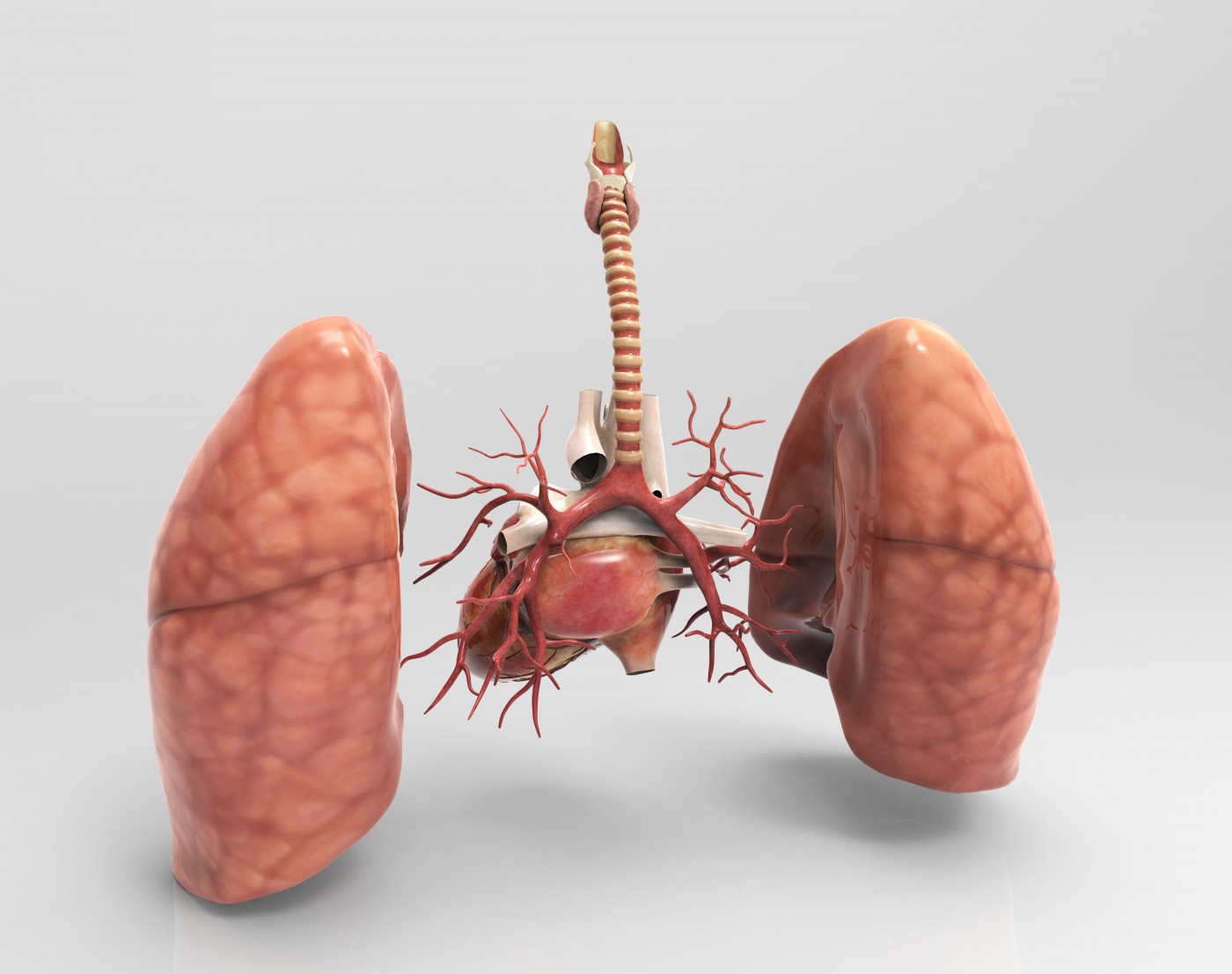
Pulmonary arteries enable the flow of blood from the heart to the lungs, where the blood picks up oxygen for the body. In PAH, the pulmonary arteries contract, forcing the heart to work faster and causing high blood pressure (hypertension).
The technique that was used in this study is called two-dimensional strain echocardiography. Although originally used to assess the function of the left ventricle of the heart, in this study it was used to examine the right ventricle. The researchers wanted to know whether use of the technique could help them better understand how the disease progresses. The study included 51 adult patients over 18 years old with PAH who were evaluated from February 2007 to June 2008 in the PAH clinic of the Cleveland Clinic in Cleveland Ohio. Most of the study participants (40) were female. The measurement that they took is known as global longitudinal strain of the right venricle (GLSRV).
By using this method of clinical assessment to examine GLSRV, the researchers were able to predict both the progression of PAH, as well as death in PAH patients.
In their report, the investigators stated “In conclusion, GLSRV showed significant correlations with conventional echocardiographic parameters of the RV systolic function. Lower absolute GLSRV is associated with the presence of adverse clinical events and deaths in patients with PAH.”
This measurement, along with GLSRV may become a standard clinical tool for assessing PAH. It could also help in assessing new treatments for PAH. The researchers noted “it will be critical to test whether it can serve as a surrogate endpoint for novel therapies in PAH. If improvement in GLSRV with vasodilator or other intervention identifies those with better clinical outcomes, then drug development in PAH could be accelerated.”
Trials of new PAH may be advanced with the use of two-dimensional strain echocardiography and GLSRV assessment.


