Pulmonary Hypertension Detection Aided By Non-invasive Doppler Echocardiography in New Study


Non-invasive techniques to detect pulmonary hypertension in individuals who are at a high risk for developing the disease are in high demand to enable more patients to be diagnosed and treated in the early stages. As the gold standard, right heart catheterization has been the primary mode of detecting an increase in pulmonary artery pressure, but other less invasive techniques such as transthoracic Doppler echocardiography may be useful to detect high pressure as well. A study from Nanjing University School of Medicine tested Doppler echocardiography in patients with interstitial lung disease and found that the technique is feasible.
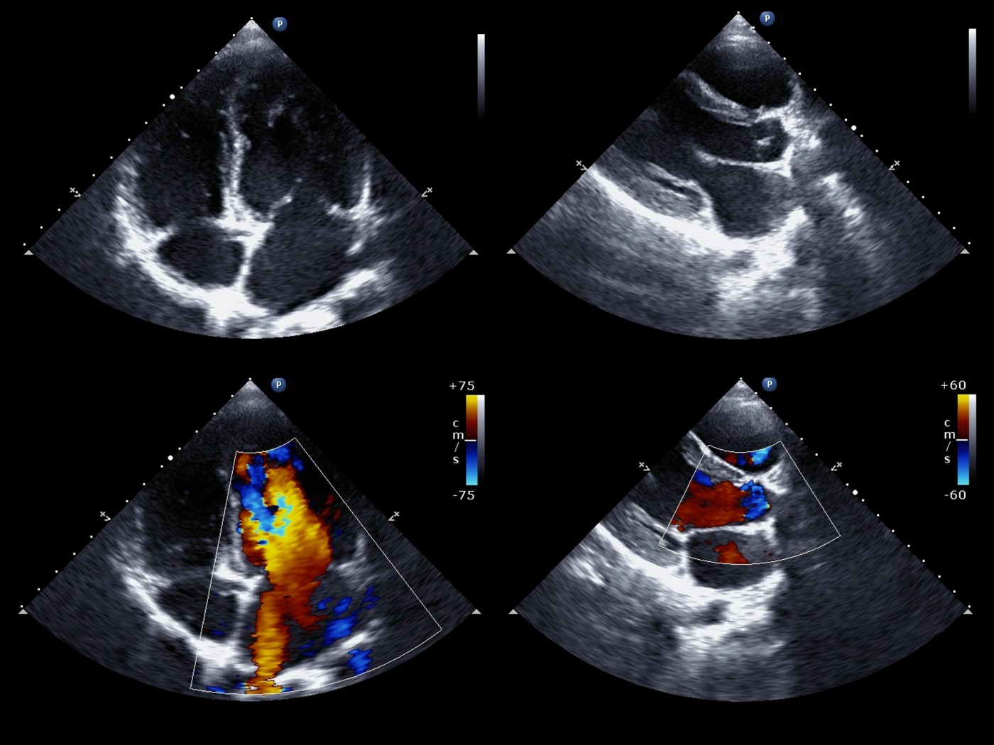
“Transthoracic echocardiography is a useful technique for noninvasive estimation of pulmonary artery pressure through tricuspid regurgitation or pulmonary valve regurgitation and is readily available anywhere,” stated the authors of “B-Lines in Assessment of Pulmonary Hypertension in Patients With Interstitial Lung Diseases: Feasibility of Transthoracic Lung Sonographic Signs,” which was published in Journal of Ultrasound in Medicine. Essentially, when blood flows through the heart valves, the fluid motion is able to be detected by Doppler echocardiography and can be analyzed for abnormalities due to pressure gradients. However, if patients do not exhibit regurgitation, an alternate method of detection is needed.
As an added feature of Doppler echocardiography, well-defined “B-lines” appear in the image as a result of interstitial lung disease. Since these B-lines are common in patients with interstitial lung disease, the researchers were interested in determining if there was a relationship between B-lines and pulmonary artery pressure.
To conduct the study, the researchers identified 134 patients with interstitial lung disease and confirmed pulmonary hypertension. They used Doppler echocardiography to see if the presence of B-lines and the distance between the lines related to an increased pulmonary artery pressure. Significantly more B-lines were present in patients with severe pulmonary hypertension, and the researchers were able to create a model to predict pulmonary artery pressure based on the number of B-lines present. Patients with more than four B-lines were almost guaranteed to have a high pulmonary artery pressure.
In the future, clinicians may be able to use B-line quantification via Doppler echocardiography in patients who do not show regurgitation to detect pulmonary hypertension. The number of B-lines can be substituted into an equation to predict pulmonary artery pressure, which can then suggest the presence of pulmonary hypertension.