MRI Can Help with PAH Evaluation and Treatment, Study Finds
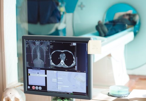


Researchers have shown that assessing vasculature structures by magnetic resonance imaging (MRI) can retrieve important information and has predictive potential for pulmonary arterial hypertension (PAH) patients.
In combination with clinical data, MRI can increase the accuracy of determining PAH severity and outcomes, and improve decisions regarding therapies.
The finding, from researchers in Sheffield, England, was reported in a study titled “Magnetic resonance imaging in the prognostic evaluation of patients with pulmonary arterial hypertension” and published in the American Journal of Respiratory and Critical Care Medicine.
Although many efforts have been made to improve PAH patients’ diagnosis and treatment, it is still a disease with a poor outcome. An accurate disease assessment and the identification of prognostic markers can help clinicians when they make treatment decisions and improve patient care.
It is common in PAH that changes in pulmonary vasculature lead to alterations in the right ventricular response with a reduction in cardiac output. This happens in the majority of cases responsible for reduced breathing and exercise capacity.
Several measurements have been used to assess disease severity and estimate prognosis, including impairment of right ventricular (RV) function, measurements of exercise capacity, and maximal oxygen uptake. However, these approaches have limitations that can lead to subjective outcome evaluations.
The use of MRI can provide an accurate and reproducible way to gather information on cardiac morphology and function. In addition, it is an imaging method that allows the identification of pulmonary vasculature with enough sensitivity to detect abnormalities or physical changes.
Researches in this study aimed to determine the value of MRI evaluation for outcome prediction in PAH patients.
They followed 576 patients with diagnosed PAH according to the ASPIRE registry who underwent MRI evaluation. The study group included 260 patients with idiopathic pulmonary arterial hypertension (IPAH) and 195 patients with PAH associated with connective tissue disease (PAH-CTD). From the total studied, 38% of the patients died.
The authors found that MRI determined parameters reflecting RV structure and stiffness of the pulmonary vasculature, specifically RV-end systolic-volume-index and relative area change of the pulmonary artery, were predictive of poorer PAH patient outcome.
MRI evaluation alone represented a three-year predictive accuracy of about 74% in the total studied population, 82% in the IPAH subgroup, and 69% predictive accuracy for the PAH-CTD subgroup.
These findings suggested that MRI results can have the same prognostic potential as the six-minute walk test (6MWT), which is associated with an outcome predictive accuracy of about 69%.
“Combining MRI measures of RV function and pulmonary artery stiffness with clinical data further improves prognostication in patients with pulmonary arterial hypertension,” the authors concluded in their report.