
VMS Heart Analysis System Accurately Evaluates Right Ventricle Remodeling in PH Patients, Study Shows
Written by |
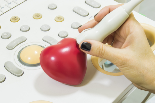
Ventripoint Diagnostics‘ VMS Heart Analysis System, which produces a two-dimensional (2D) volumetric reconstruction model of the heart, accurately measures small changes in the heart’s right ventricle volume and can be used in patients with pulmonary hypertension (PH), a new study finds.
The study, “Two-dimensional knowledge-based volumetric reconstruction of the right ventricle documents short-term improvement in pulmonary hypertension,” appeared in the journal Echocardiography.
PH is characterized by increased blood pressure of the right ventricle. This leads to expansion of the ventricle and its eventual failure. Doctors use 2D echocardiography as the primary imaging test to diagnose PH, though its limitations can significantly affect the clinical evaluation of a patient’s condition.
For this reason, cardiac magnetic resonance (CMR) imaging has become the gold standard for right-ventricle imaging. Yet this method is expensive, resource-consuming and unavailable for many patients.
Recognizing the need for an affordable, accurate and reliable right-ventricle assessment imaging method, a research team at England’s Royal Free Hospital in London compared the diagnosis efficacy of the new VMS Heart Analysis System with the standard CMR method.
They evaluated 25 patients with pulmonary arterial hypertension (PAH), 15 of whom had connective tissue disease (CTD)-associated PAH, and six with chronic thromboembolic pulmonary hypertension.
Researchers found they could use the 2D VMS Heart Analysis System instead of cardiac MRI to measure alterations in the right ventricle over time.
“Ventricular remodeling in PAH can be differentiated into two patterns: adaptive remodeling with concentric hypertrophy and preserved function, and maladaptive remodeling with eccentric hypertrophy and worsening function,” researchers wrote. “Our study shows that within several months a change from one pattern to the other can occur with medical therapy, even in CTD-associated PAH.”
Having ways to monitor the clinical remodeling progress of the right ventricle in PH patients is critical in determining the efficacy of various therapeutics. The VMS System showed it could predict patient outcomes by detecting right-ventricle volume reduction in those who responded to treatment, and increased volume in those with worse clinical outcomes.
The team concluded that the 2D VMS Heart Analysis System “can be reliably used in a busy clinical setting to follow up right-ventricular indices in pulmonary hypertension,” and that the system “is a feasible substitute for cardiac MRI.”


Leave a comment
Fill in the required fields to post. Your email address will not be published.