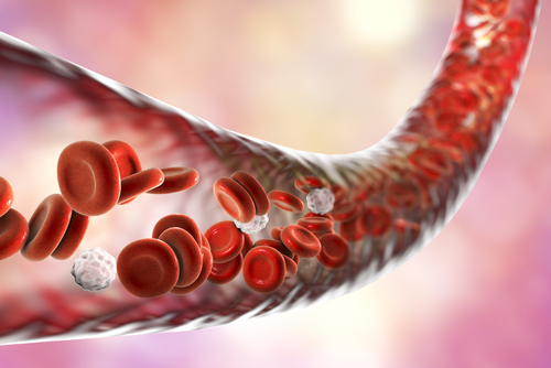
Increased Respiratory Capacity Found in Platelets of Group 2 PH Patients

Patients with group 2 pulmonary hypertension — those with PH due to left heart disease — show increased platelet respiration, which is associated with worse right ventricular function, according to new research.
The study, “Alterations in platelet bioenergetics in Group 2 PH-HFpEF patients,” was published in the journal PLOS ONE.
Patients with group 1 pulmonary arterial hypertension (PAH) have altered energy metabolism in the vascular cells of the lungs, and in cardiac and skeletal muscle, which is in turn associated with vascular remodeling, heart failure, and exercise intolerance.
Scientists at Pennsylvania’s University of Pittsburgh also showed that these patients’ platelets (blood cells involved in clotting) mirror such changes, as illustrated by their increased glycolysis — the breakdown of glucose (sugar) to produce energy — due to a switch in energy source that favors a greater oxidation of fatty acids in mitochondria, the cells’ power plants.
Yet whether such changes also occur in other PH subgroups, including the most prevalent subtype, group 2, remained to be determined.
Aiming to address this gap, the team at Pittsburgh extracted platelets from 20 patients with group 2 PH (mean age 69.4 years, 10 women) with heart failure but preserved ejection fraction — how much blood the heart pumps out with each contraction — and 20 controls to measure platelet bioenergetics and hemodynamic (blood flow) parameters.
Of note, the PH patients analyzed had participated in a Phase 2 trial (NCT01431313) testing whether inhaled nitrite — a vasodilator shown to improve blood flow dynamics and to regulate mitochondrial function in patients with group 1 and group 2 PH — altered pulmonary vascular resistance, an indicator of disease severity.
The results showed that, unlike group 1 PH patients, the platelets of those with group 2 PH do not have altered glycolysis compared with the controls. However, the cells’ maximal capacity of respiration (which takes place within mitochondria) — the oxygen consumption rate and the reserve respiratory capacity — was increased due to an increase in fatty acid oxidation and, to a lesser degree, to higher glucose oxidation.
Inhaled nitrite did not alter this increased maximal capacity of cellular respiration, despite changes in blood flow “arguing further that the mitochondrial changes in group 2 PH are likely not completely secondary to hemodynamic changes,” the researchers said.
The data also showed that the higher this maximal capacity, the lower the right ventricle (RV) stroke work index, a marker of RV function. RV function is a known predictor of outcomes in patients with PH and other cardiovascular diseases.
“We demonstrate that unlike Group 1 PH, platelets from Group 2 PH patients do not show a change in glycolytic rate compared to healthy controls. However, they did show a significant increase in maximal respiratory capacity similar to Group 1 subjects. Notably, this enhanced maximal respiration negatively correlated with patient RV hemodynamics,” the researchers stated.
Though not statistically significant, the data revealed a trend toward higher reserve oxygen consumption rate being correlated with higher body mass index and with diabetes (found in 13 patients), “thus linking platelet metabolism to diabetes and insulin resistance,” the researchers said.
Overall, the data “demonstrate that Group 2 PH subjects have altered bioenergetic function, though this alteration is not identical to that of Group 1 PH,” the team concluded.
“These data start to distinguish the metabolic phenotypes of Group 1 and Group 2 PH, and may have important implications for delineating the hemodynamic versus non-hemodynamic modulation of mitochondrial function,” they added.


