
Researchers Advance Pulmonary Embolism Diagnosis Through Computed Tomography
Written by |
Researchers at Leiden University Medical Center in The Netherlands recently published in the journal Diagnostic and Interventional Radiology a review on the value of computed tomography (CT) as a diagnostic tool for pulmonary embolism. The study is entitled “The role of computed tomography in the diagnosis of acute and chronic pulmonary embolism.”
Acute pulmonary embolism is a life-threatening cardiovascular condition in which blood clots block one of the pulmonary arteries in the lungs, possibly leading to sudden breathing problems, rapid heart and breathing rates, cough with blood, chest pain and fainting. The prognosis of the condition mainly depends on the severity of right ventricular dysfunction and on residual pulmonary circulation.
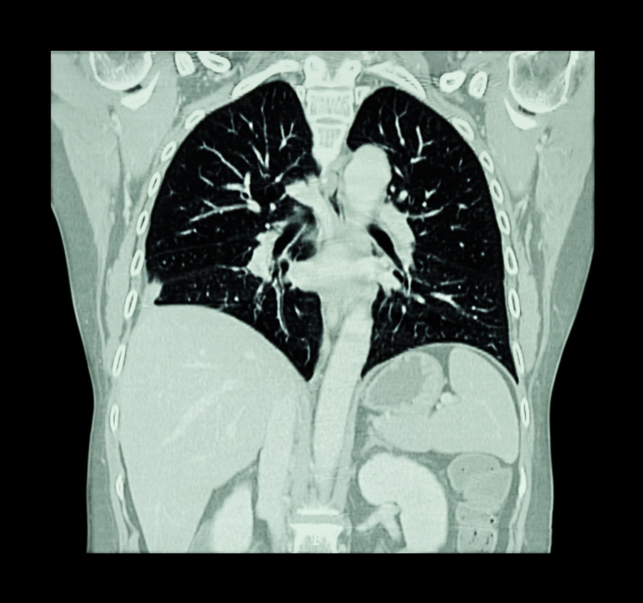
According to the authors, the diagnosis of acute pulmonary embolism primarily relies on non-invasive imaging techniques, namely through the fast, accurate, highly specific and widely available computed tomography pulmonary angiography (CTPA). However, CTPA tends to be overused, which may result in an unnecessary risk for malignancies due to the patient exposure to X-rays. A correct implementation of clinical decision rules in the diagnosis of pulmonary embolism is needed to improve the adequate use of the technique.
The team reports that CTPA is an important resource to include or exclude a pulmonary embolism diagnosis. CT is also a prognostic tool as it evaluates the patient’s right ventricular function. In this case, a simple ratio between right ventricular and left ventricular diameter that is higher than 1.0 indicates a risk for adverse outcome, while a ratio lower than 1.0 can safely exclude adverse outcome.
Upon experiencing acute pulmonary embolism, a few patients can develop chronic thromboembolic pulmonary hypertension (CTEPH), a life-threatening condition caused by repeated or unresolved pulmonary embolism that can lead to an abnormally high blood pressure in the pulmonary arteries — pulmonary hypertension. CTEPH patients can experience shortness of breath during exercise, chest discomfort and fatigue. The condition is a major cause of severe pulmonary hypertension and right heart failure.
CTEPH can be treated through a surgical procedure called thrombectomy that allows the removal of the blood clots. According to the study, CT can also diagnose CTEPH and is becoming the imaging method of choice for CTEPH diagnosis and in the identification of patients who might benefit from thrombectomy.
The team suggests that new developments and refinement in the CT field may further increase its value as a diagnostic and prognostic tool in order to improve patient care for those with CTEPH or pulmonary embolisms.


Leave a comment
Fill in the required fields to post. Your email address will not be published.