
Same Procedure, Two Outcomes for Right Ventricular Failure in PH
Written by |
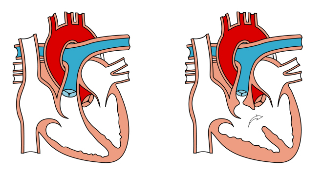
The right ventricle is extremely critical to the progression of pulmonary hypertension due to its position immediately before the pulmonary artery. It was previously shown the volume of the right ventricle can predict changes in clinically stable pulmonary hypertension, and now scientists know that different severities of right ventricular failure (RVF) due to chronic pulmonary artery banding in rats leads to different clinical characteristics.
A research team at Beatrix Children’s Hospital spearheaded the investigation “Clinical Symptoms of Right Ventricular Failure in Experimental Chronic Pressure Load are Associated with Progressive Diastolic Dysfunction.” To begin, the researchers placed a band around the pulmonary artery to induce RVF in 12 rats and left 7 rats in a healthy state. Within 52 days, five of the 12 rats developed symptoms of RVF.
[adrotate group=”4″]
Between the symptom-free and symptomatic pulmonary artery banding groups, diastolic function was poorer with symptoms, and contractility was better in the asymptomatic rats. Cardiac output and right ventricular stroke volume were reduced in either group, and end diastolic pressure was increased.
As part of the mechanism of RVF progression, fatty acids and carbohydrates may play a role. The researchers found downregulation of both fatty acid oxidation and carbohydrate metabolism in the symptomatic rats.
[adrotate group=”3″]
Overall, rats more prone to hypoxia may have been more susceptible to RVF. Diastolic function deteriorated with chronic pulmonary artery banding, and severe clinical symptoms appeared. Future efforts by this group and others may use this model of RVF to investigate the impact of therapeutic interventions on RVF.


Leave a comment
Fill in the required fields to post. Your email address will not be published.