Worse PH Prognosis Linked to Exercise-induced Increase in Arterial Pressure, Study Suggests

An increase in right atrial blood pressure during exercise in people with pulmonary hypertension (PH) reflects problems with blood flow in the heart and is linked to a worse prognosis and higher mortality, a study shows.
Researchers therefore suggest that right atrial blood pressure during exercise should be considered an essential prognostic indicator in these patients.
The study, “Right Atrial Pressure During Exercise Predicts Survival in Patients With Pulmonary Hypertension,” was published in the Journal of the American Heart Association.
Studies have demonstrated that an elevated right atrial blood pressure (RAP) when resting, as measured by right heart catheterization, is associated with a poor PH prognosis.
However, little is known about RAP during exercise in PH and its value as a prognostic tool.
To investigate further, researchers at the University Hospital Zürich in Switzerland analyzed medical records of 270 people who had right heart catheterization during exercise and evaluated its prognostic value by assessing patient outcomes over a median observation period of 3.7 years.
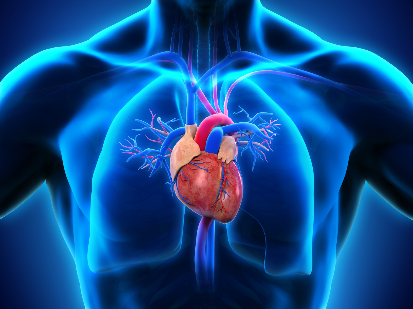
Of note, right heart catheterization (RHC) measures blood pressure in the right atrium of the heart and in the arteries leading to the lungs (pulmonary artery pressure or PAP) by inserting a flexible tube through blood vessels into the right side of the heart and pulmonary arteries.
The median (middle) age of those selected was 62 years, and the group included 63% women.
In total, 152 people had resting PH (25 mmHg mean PAP or more) and 58 had PH induced by exercise (resting mean PAP less than 25 mmHg, exercise mean greater than 30 mmHg). For comparison, 60 subjects without PH were assessed.
Exercise was performed using a cycling machine while participants laid on their backs. PAP and RAP pressure was measured continuously and averaged over several breathing cycles after at least 15 minutes of rest and in the last 30 seconds of exercise.
Results showed that the median peak workload (power) in those without PH was 40 watts, while people with exercise-induced PH managed 30 watts, and patients with PH 20 watts.
RAP decreased or was unchanged in non-PH subjects (6 mmHg) but increased during exercise in both PH (8 to 13 mmHg at peak exercise) and exercise-induced PH patients (6 to 9 mmHg at peak exercise), with the highest RAP at the end of exercise measured in patients with PH.
The mean PAP increased in all groups during exercise, with the highest change seen in patients with PH (34 to 54 mmHg) and those with exercise-induced PH (21 to 37 mmHg).
The heart rate and stroke volume (volume of blood pumped from the left ventricle per beat) and related measures of cardiac output (amount of blood pumped through the circulatory system), and cardiac index (heart performance based on subject body size) increased with exercise in all groups, being significantly higher in healthy subjects compared with PH patients.
As determined by the mean PAP to cardiac output ratio, the pressure to blood flow relationship did not change in healthy participants but increased in exercise-induced PH and even more in PH patients. In contrast, the RAP to cardiac output relationship significantly decreased in those without PH, while significantly increasing in exercise-induced PH and PH groups.
Compared with patients with unchanged or decreasing RAP during exercise, those with an exercise‐associated increase in RAP had a greater than fourfold increase in the risk of death or lung transplant, and also a shorter transplant‐free survival — 8.3 vs. 7.0 years.
A statistical analysis found that an exercise‐induced increase in RAP was a significant predictor of reduced transplant‐free survival even after adjusting for age, resting RAP, and heart rate.
Additional predictors of transplant‐free survival were age, diagnostic group, New York Heart Association functional class (heart failure classification), and the distance walked in six minutes (6MWD), with younger age and higher 6MWD being the strongest significant predictors.
At rest, low heart rate and adequate arterial blood oxygenation were significant predictors of transplant‐free survival, while during exercise, changes in all measures of blood flow were predictors except for arterial blood oxygenation.
Changes in RAP induced by exercise were significantly linked with several clinical and blood flow variables, with mean PAP to cardiac output ratio being the best single predictor of transplant‐free survival.
“RAP increase during exercise, as observed in [exercise-induced PH] and PH, reflects [blood flow] impairment and poor prognosis,” the researchers concluded.
“Therefore, our data suggest that changes in RAP during exercise right heart catheterization are clinically important indexes of the cardiovascular function,” they added.


